


주메뉴
- About IBS 연구원소개
-
Research Centers
연구단소개
- Research Outcomes
- Mathematics
- Physics
- Center for Theoretical Physics of the Universe(Particle Theory and Cosmology Group)
- Center for Theoretical Physics of the Universe(Cosmology, Gravity and Astroparticle Physics Group)
- Center for Exotic Nuclear Studies
- Center for Artificial Low Dimensional Electronic Systems
- Center for Underground Physics
- Center for Axion and Precision Physics Research
- Center for Theoretical Physics of Complex Systems
- Center for Quantum Nanoscience
- Center for Van der Waals Quantum Solids
- Chemistry
- Life Sciences
- Earth Science
- Interdisciplinary
- Institutes
- Korea Virus Research Institute
- News Center 뉴스 센터
- Career 인재초빙
- Living in Korea IBS School-UST
- IBS School 윤리경영

주메뉴
- About IBS
-
Research Centers
- Research Outcomes
- Mathematics
- Physics
- Center for Theoretical Physics of the Universe(Particle Theory and Cosmology Group)
- Center for Theoretical Physics of the Universe(Cosmology, Gravity and Astroparticle Physics Group)
- Center for Exotic Nuclear Studies
- Center for Artificial Low Dimensional Electronic Systems
- Center for Underground Physics
- Center for Axion and Precision Physics Research
- Center for Theoretical Physics of Complex Systems
- Center for Quantum Nanoscience
- Center for Van der Waals Quantum Solids
- Chemistry
- Life Sciences
- Earth Science
- Interdisciplinary
- Institutes
- Korea Virus Research Institute
- News Center
- Career
- Living in Korea
- IBS School


News Center

| Title | New Study Reveals How the Brain Integrates Pain Prediction and Stimuli | ||
|---|---|---|---|
| Embargo date | 2024-09-23 16:33 | Hits | 22 |
| Press release |
 New Study Reveals How the Brain Integrates Pain Prediction and Stimuli.docx
|
||
| att. | |||
New Study Reveals How the Brain Integrates Pain Prediction and Stimuli- Uncovering the neural mechanisms of pain processing using functional Magnetic Resonance Imaging (fMRI) - A study led by Associate Director WOO Choong-Wan of the Center for Neuroscience Imaging Research (CNIR) within the Institute for Basic Science (IBS), along with Michael YOO Seng Bum, Assistant Professor of Biomedical Engineering at Sungkyunkwan University, has uncovered new insights into how the brain processes and integrates pain information. Their research goes beyond identifying brain areas that respond to pain, revealing the mechanisms behind the brain's integration of pain-related information. Using functional magnetic resonance imaging (fMRI), they formalized how the brain combines pain expectations with the actual intensity of painful stimuli. Pain is a complex experience influenced not just by the intensity of a painful stimulus but also by the individual’s expectations. For instance, the pain one expects to feel can alter the perception of the actual pain experienced. While previous research has mapped out which brain regions handle these separate factors that contribute to our pain experience, this new study tackles the question of how these different factors come together to create a unified sensation of pain. KIM Jungwoo, the first author of the study, stated, “It’s not just about knowing which parts of the brain are important; ultimately, understanding how pain arises is key to figuring out how to eliminate unnecessary pain.” The researchers used fMRI to observe brain activity in participants exposed to varying levels of pain stimuli, while also manipulating their expectations about the level of pain they would feel. To fully understand how pain is processed in the brain, they separated the process into two stages: preservation (how the brain maintains information about pain expectations and stimulus intensity) and integration (how these elements combine to form a cohesive pain experience). They examined these processes across different levels of the brain’s cortical hierarchy*, expecting lower-level brain networks to preserve information without integrating it, and higher-level networks to preserve and integrate both. * Cortical Hierarchy: The brain processes information in a stepwise manner, with lower-level networks (like the sensory and motor networks) handling basic sensory input, and higher-level networks (such as the limbic system and default mode network) integrating more complex information. This study used this framework to understand how the brain processes and integrates pain information at different levels. Contrary to the researchers’ initial hypothesis, the results showed that all networks, regardless of level, preserved both types of information—pain expectations and stimulus intensity. However, only higher-level networks were able to integrate this information by simply adding the preserved expectation and stimulus information together. This suggests that while the entire brain stores pain information, only specific areas are responsible for integrating different pain-related signals into the experience of pain. This study represents a significant collaboration between two fields of neuroscience. Dr. Yoo, an expert in decision-making and electrophysiology, and Dr. Woo, a pain researcher specializing in fMRI, combined their expertise to explore how pain information is processed across the whole brain. Their innovative approach sheds light on the brain’s mechanisms for processing pain, providing valuable insights that could lead to new approaches to treating chronic pain. Michael YOO Seng Bum, the co-lead author said “It was a meaningful collaborative study that combined the strengths of each principal investigator to advance beyond merely reporting the activation of specific regions, allowing us to investigate principles of how information is integrated across the brain.” WOO Choong-Wan, another co-lead author, described the research as “an innovative study using geometric information encoded in brain activation patterns to reveal the integration mechanism of distinct types of pain information,” adding that “this discovery would not have been possible without a collaboration.”
Notes for editors
- References
- Media Contact
- About the Institute for Basic Science (IBS)
|
|||
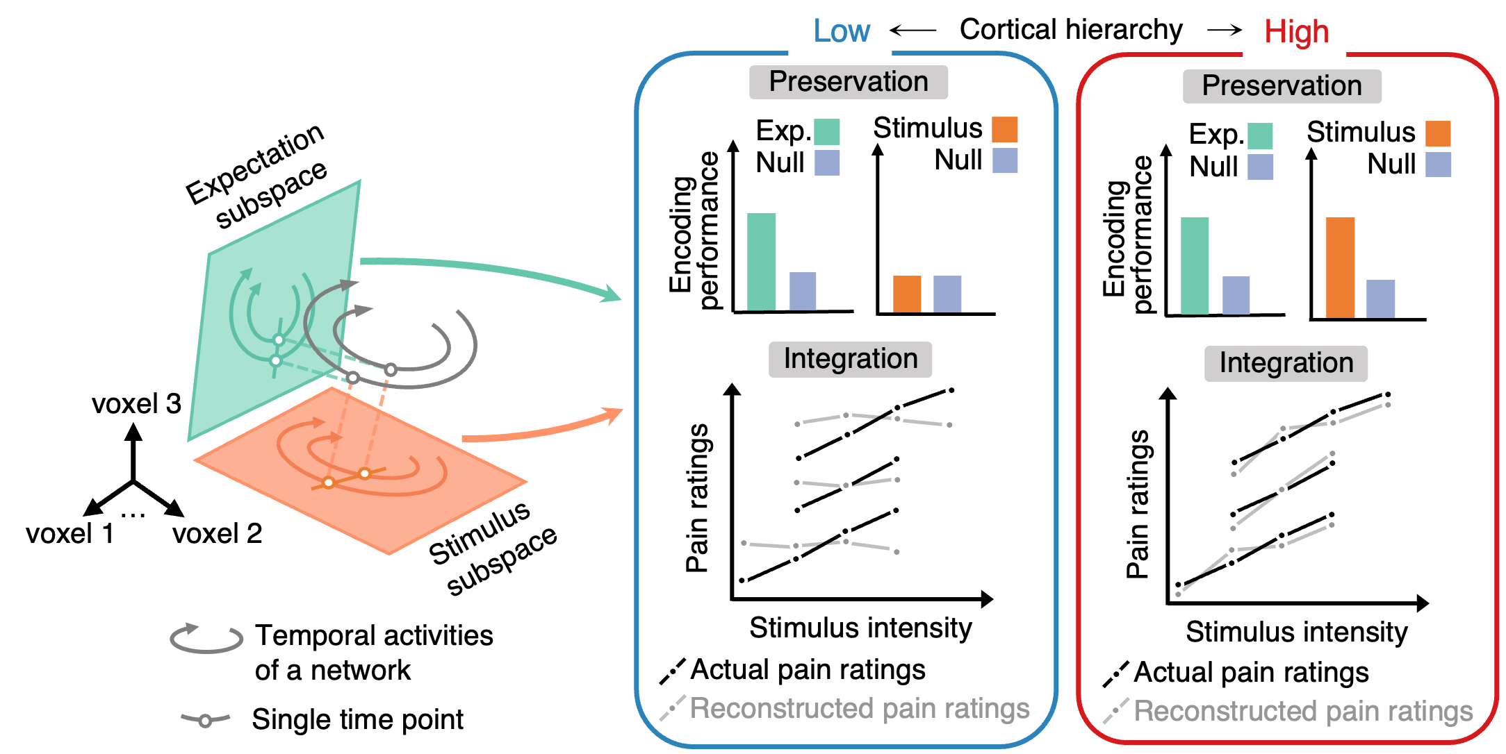 Figure 1. Hypothesis on the preservation and integration of pain information
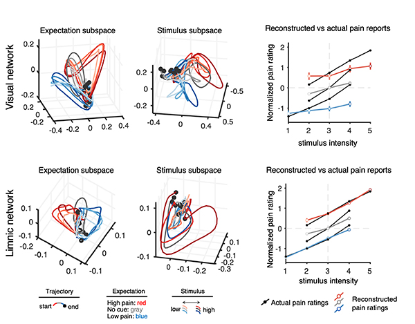 Figure 2. Subspace patterns and comparison of reconstructed vs. actual pain reports
| Next | |
|---|---|
| before |
- Content Manager
- Communications Team : Kwon Ye Seul 042-878-8237
- Last Update 2023-11-28 14:20
55, Expo-ro, Yuseong-gu, Daejeon, Korea, 34126
Tel. +82-42-878-8114 | Fax. +82-42-878-8079 | E-mail. webmaster@ibs.re.kr
Copyright(c) IBS. All Rights Reserved.
