주메뉴
- About IBS 연구원소개
-
Research Centers
연구단소개
- Research Outcomes
- Mathematics
- Physics
- Center for Theoretical Physics of the Universe(Particle Theory and Cosmology Group)
- Center for Theoretical Physics of the Universe(Cosmology, Gravity and Astroparticle Physics Group)
- Center for Exotic Nuclear Studies
- Center for Artificial Low Dimensional Electronic Systems
- Center for Underground Physics
- Center for Axion and Precision Physics Research
- Center for Theoretical Physics of Complex Systems
- Center for Quantum Nanoscience
- Center for Van der Waals Quantum Solids
- Chemistry
- Life Sciences
- Earth Science
- Interdisciplinary
- Center for Neuroscience Imaging Research(Neuro Technology Group)
- Center for Neuroscience Imaging Research(Cognitive and Computational Neuroscience Group)
- Center for Algorithmic and Robotized Synthesis
- Center for Genome Engineering
- Center for Nanomedicine
- Center for Biomolecular and Cellular Structure
- Center for 2D Quantum Heterostructures
- Center for Quantum Conversion Research
- Institutes
- Korea Virus Research Institute
- News Center 뉴스 센터
- Career 인재초빙
- Living in Korea IBS School-UST
- IBS School 윤리경영


주메뉴
- About IBS
-
Research Centers
- Research Outcomes
- Mathematics
- Physics
- Center for Theoretical Physics of the Universe(Particle Theory and Cosmology Group)
- Center for Theoretical Physics of the Universe(Cosmology, Gravity and Astroparticle Physics Group)
- Center for Exotic Nuclear Studies
- Center for Artificial Low Dimensional Electronic Systems
- Center for Underground Physics
- Center for Axion and Precision Physics Research
- Center for Theoretical Physics of Complex Systems
- Center for Quantum Nanoscience
- Center for Van der Waals Quantum Solids
- Chemistry
- Life Sciences
- Earth Science
- Interdisciplinary
- Center for Neuroscience Imaging Research(Neuro Technology Group)
- Center for Neuroscience Imaging Research(Cognitive and Computational Neuroscience Group)
- Center for Algorithmic and Robotized Synthesis
- Center for Genome Engineering
- Center for Nanomedicine
- Center for Biomolecular and Cellular Structure
- Center for 2D Quantum Heterostructures
- Center for Quantum Conversion Research
- Institutes
- Korea Virus Research Institute
- News Center
- Career
- Living in Korea
- IBS School
News Center
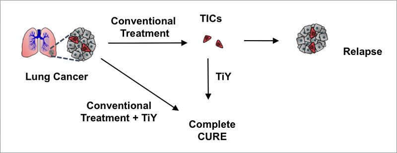
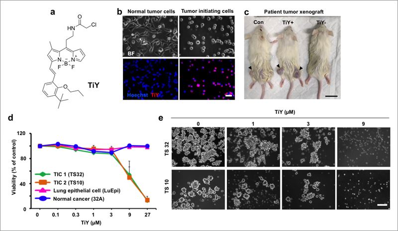
Versatile Sensor Against Tumor Initiating Cells- New sensor identifies and suppresses tumor initiating cells, Most cancer deaths are caused by recurrent or metastatic tumors. Conventional therapies target rapidly dividing tumor cells, but are unable to eradicate the highly chemoresistant tumor initiating cells (TICs), ultimately responsible for relapse and spreading of the tumors in other parts of the body. A team of researchers at the Center for Self-assembly and Complexity, within the Institute for Basic Science (IBS) developed the first fluorescent sensor to visualize TICs. Functional in lung, central nervous system, melanoma, breast, renal, ovarian, colon, and prostate cancer cell cultures, this could become a useful tool for biopsy-free post-treatment assessment and anti-TIC drug development. The study was conducted in POSTECH (Pohang, South Korea), in collaboration with the Agency for Science Technology and Research (A*STAR, Singapore), and is published in Angewandte Chemie International Edition. In some cases, a minority of cells called TICs, or cancer stem cells, are culpable of repopulating the tumor after therapy. Their stem cell-like properties enable them to maintain a pool of cancer stem cells within the tumor, as well as to produce new mature tumor cells. Although some TIC-targeting antibodies are available, there is not a single universal antibody which covers all kinds of TICs from different tissues. By screening thousands of fluorescent chemicals, the team developed TiY, a sensor that selectively stained the TICs of non-small cell lung cancer, which accounts for around 85% of all lung cancers. They found out that TiY is capable of distinguishing TICs from non-TICs in various human lung cancer cell lines and patient-derived lung tumors.
Beyond lung cancer, TiY is able to target TICs in 28 types of human cell lines derived from the central nervous system, melanoma, breast, renal, ovarian, colon, and prostate cancer. “TiY has the features of a universal probe, applicable to various kinds of cancer regardless of the origin of the tissue,” explains CHANG Young-Tae.
The researchers have discovered that TiY binds to an intracellular protein called vimentin. Part of the cytoskeleton, vimentin gives flexibility and structure to the cell, but it is also a marker of epithelial-mesenchymal transition (EMT), which is considered a crucial event for metastasis in epithelial tumors. The EMT process allows cells to detach from their neighbors and adopt a migratory and invasive behavior. Although this is a normal process in embryogenesis to build new tissues, when it occurs in cancer cells it generates metastasis. Although vimentin is a known TIC biomarker and anti-vimentin antibodies are available, these cannot enter living cells, and thus could not be used for live TIC detection. On the contrary, TiY is a drug-like small molecule with the unique property of detecting TICs in vivo without biopsy and isolating viable TICs for further studies. In the lab, TICs are able to grow as sphere-looking structures, the so-called tumor spheres, easily recognizable under the microscope. The researchers have discovered that this sphere-forming ability is directly influenced by vimentin as higher concentrations of TiY resulted in the inhibition of sphere formation. Moreover, the team compared TiY with a known vimentin-inhibitor, withaferin A (WFA) and observed that TiY has a stronger selectivity towards TICs than WFA, when compared with toxicity to normal cells and non-TIC cells.
Additional experiments also showed that TiY can suppress the tumor growth in mice xenograft model. Currently, studies for comprehensive application of TiY to a broader range of cancer examples are underway, together with screenings of anti-TIC drug platforms.
Letizia Diamante Notes for editors - References - Media Contact - About the Institute for Basic Science (IBS) |
|||
Center for Self-assembly and ComplexityPublication Repository |
|||
|
|
| Next | |
|---|---|
| before |
- Content Manager
- Public Relations Team : Yim Ji Yeob 042-878-8173
- Last Update 2023-11-28 14:20


